Insights into all the latest

Research in the News

Tracking the pulse of the latest spinal cord injury research news — from lab breakthroughs to clinical milestones.

Author's Current Top 3 to Read

1

10 MAY 2026

Could Skin-Level Spinal Cord Stimulation Help Calm Autonomic Dysreflexia After SCI?

A new UBC thesis study in rats suggests that transcutaneous spinal cord stimulation may reduce autonomic dysreflexia by activating the spinal cord's own inhibitory 'braking system', helping quiet the overactive reflexes that drive dangerous blood pressure spikes after high-level SCI.

Deep Dive

Autonomic dysreflexia, often shortened to AD, is one of the most frightening secondary complications of spinal cord injury.

It can come on fast. A blocked catheter, full bladder, bowel problem, skin irritation, pressure sore, tight clothing, or another trigger below the injury level can cause a sudden spike in blood pressure. For people with cervical or upper thoracic SCI, this can mean pounding headache, sweating, flushing, goosebumps, anxiety, chest tightness, slow heart rate, and a real medical emergency.

This story is especially relevant for people who live with severe or repeated AD. The study does not offer an immediate home treatment, but it helps explain why non-invasive spinal cord stimulation could become a useful tool for reducing AD in the future.

The research comes from a 2026 University of British Columbia thesis by Hari Prasad Joshi, supervised by Professor Andrei Krassioukov, a leading researcher in autonomic dysfunction after spinal cord injury.

Author(s):

Hari Prasad Joshi. Supervisory and examining contributors included Andrei V. Krassioukov, Ismail Laher, Rahul Sachdeva, Soshi Samejima, Alexander G. Rabchevsky, and Keith E. Tansey.

Source:

University of British Columbia, Faculty of Graduate and Postdoctoral Studies, Experimental Medicine. Thesis title: "Unraveling the Mechanism of Transcutaneous Spinal Cord Stimulation in Mitigating Autonomic Dysreflexia in Experimental Spinal Cord Injury."

What AD is, in plain English

The autonomic nervous system controls automatic jobs: blood pressure, heart rate, sweating, blood vessel tightening, bladder, bowel, temperature regulation, and more.

After a high spinal cord injury, the brain can lose proper control over many of these automatic circuits below the injury. The lower body can still send alarm signals into the spinal cord, but the brain's calming control cannot always get back down properly.

A useful analogy is a smoke alarm that is wired to a sprinkler system but no longer connected to the control room. Something small sets off the alarm, the sprinkler system overreacts, and nobody upstairs can easily switch it off.

In AD, a trigger below the injury level can cause blood vessels below the injury to clamp down hard. Blood pressure rises quickly. The brain senses the high pressure and tries to slow the heart, but it cannot fully relax the blood vessels below the injury because the spinal cord pathway is interrupted.

That is why AD is not just uncomfortable. It can be dangerous.

The idea: use stimulation to restore the spinal cord's brake

The study looked at transcutaneous spinal cord stimulation, or tSCS.

"Transcutaneous" means through the skin. Unlike implanted epidural stimulation, tSCS uses surface electrodes placed on the skin over the spine. The goal is to send electrical pulses into spinal circuits without surgery.

In this rat study, stimulation was delivered over the T7 spinal segment using 30 Hz pulses for 60 minutes. The model involved a high thoracic T3 spinal cord contusion injury. Six weeks later, the researchers triggered AD-like spinal activity using colorectal distension, a standard experimental model because bowel or rectal distension is a common real-world AD trigger.

The big question was not simply "does stimulation reduce AD?" Previous work had already suggested tSCS can reduce AD. This study asked a deeper question: how might it work inside the spinal cord?

The spinal cord after SCI can become too excitable

After SCI, the spinal cord below the injury is not silent. It can rewire itself.

Some rewiring may be helpful. But some becomes maladaptive, meaning it makes symptoms worse. In AD, sensory signals from the bowel, bladder, skin, or other areas below the injury can over-activate spinal circuits that control sympathetic output. Sympathetic output is the "fight or flight" side of the autonomic system, and when it overfires it can drive blood pressure dangerously high.

The key cells in this process include sympathetic preganglionic neurons, or SPNs. These are spinal neurons that help send commands to the sympathetic nervous system. When they become overactive after a trigger, they can contribute to the blood pressure surge of AD.

The thesis found that colorectal distension strongly activated spinal neurons and SPNs after SCI. That fits the AD pattern: a visceral trigger below the injury sends a powerful signal into overexcitable spinal autonomic circuits.

What stimulation changed

When tSCS was applied at the same time as colorectal distension, the overactivation was reduced.

The researchers used markers such as c-Fos to identify activated neurons. They found that the CRD trigger increased neuronal activation across multiple spinal segments. But when CRD was paired with tSCS, that activation was significantly reduced compared with CRD alone.

Most importantly, tSCS reduced activation of the sympathetic preganglionic neurons involved in driving autonomic output.

At the same time, tSCS increased activation of inhibitory spinal interneurons. These are local spinal neurons that help quiet other neurons. The study focused on inhibitory systems involving GABA and glycine, two chemical messengers that act like brakes in the nervous system.

In plain English: stimulation appeared to turn down the overactive alarm circuit and turn up the spinal cord's braking system.

Why GABA and glycine matter

GABA and glycine are inhibitory signals. They help stop nerve circuits from firing too strongly.

In everyday terms, if excitatory signals are the accelerator, inhibitory signals are the brake. AD may partly happen when the accelerator gets stuck and the brake is too weak. This study suggests tSCS may help press the brake at the spinal level.

That does not mean tSCS "cures" AD. It means stimulation may change the balance inside the spinal cord so that bowel or bladder signals are less likely to explode into a dangerous sympathetic reflex.

Why this matters for people who live with AD

For someone who gets severe AD, the practical goal is not abstract. It is fewer dangerous spikes, less fear around bladder and bowel routines, better sleep, less strain on the heart and brain, and more confidence that the body will not suddenly swing into crisis.

Repeated AD episodes may also matter for long-term health. Blood pressure instability after SCI is linked to cardiovascular and cerebrovascular risk, and researchers increasingly worry that repeated highs and lows could affect brain blood flow and cognition over time.

A non-invasive stimulation method would be especially valuable because it could potentially be used repeatedly, adjusted to the individual, and delivered without implanted surgery.

What this study does not prove yet

This was an experimental rat study, not a human clinical trial.

The researchers studied spinal cord tissue and neuronal activation patterns after stimulation. That gives important mechanism information, but it does not by itself prove that the same stimulation settings will safely and reliably prevent AD in people.

The study also used a specific injury model, timing, stimulation site, and stimulation parameters. Human SCI is more varied: injury level, completeness, time since injury, bladder and bowel routines, medications, spasticity, pain, skin triggers, and cardiovascular health all differ between people.

So the fair conclusion is careful but encouraging: tSCS appears to recruit inhibitory spinal circuits that can dampen AD-related sympathetic activation in an animal model. That gives researchers a stronger reason to test and refine tSCS for AD in people with SCI.

Why this matters in SCI

AD is often treated as a management problem: find the trigger, sit upright, loosen clothing, empty the bladder or bowel, check the skin, monitor blood pressure, and use medication if needed. That emergency approach remains essential.

But this research points toward something more preventive: changing the spinal cord circuits that make AD so explosive in the first place.

If future human studies confirm the effect, tSCS could become part of autonomic rehabilitation. Not just rehabilitation for muscles or walking, but rehabilitation for blood pressure control and internal body stability.

For people living with severe AD, that would be a meaningful goal.

Reader Q&A

Could I use a home electrical stimulator to treat AD now?

No. This should not be tried independently. AD can be dangerous, and spinal stimulation settings, electrode placement, and patient selection need clinical testing. Anyone having AD episodes should follow their medical AD plan and speak with their SCI clinician.

Is this only relevant to bowel-triggered AD?

The experiment used colorectal distension because bowel and rectal distension are reliable AD triggers in research and real life. The mechanism may also matter for bladder or other triggers, but that needs direct testing.

Why is this exciting if it was only in rats?

Because it explains a possible mechanism. It suggests tSCS may work by activating inhibitory spinal circuits, not just by generally "stimulating the spine". Mechanism matters because it helps researchers choose better stimulation locations, strengths, and timing for future human trials.

Read full article → (external link)
2

9 MAY 2026

The Brain May Move With the Abdomen: Why This Could Matter for Brain Fog and Blood Pressure Problems After SCI

A new Nature Neuroscience study shows that brain motion in awake mice is driven by mechanical coupling with the abdomen, not mainly by breathing or heartbeat. This is not an SCI study, but it may open a new way to think about secondary problems after spinal cord injury, including blood pressure instability, altered cerebral blood flow, and cognitive fatigue.

Deep Dive

This is a News + story. The main paper is not about spinal cord injury, but it may matter to SCI research because it reveals a body-brain mechanism that has been largely overlooked.

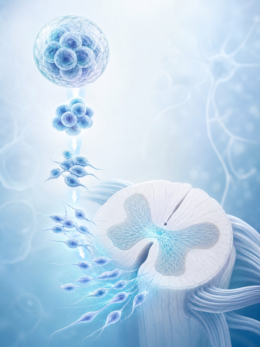
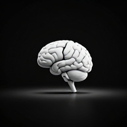
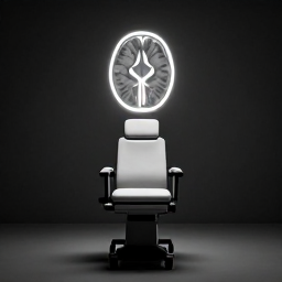
The study, published in Nature Neuroscience, found that the brain is mechanically linked to the abdomen. In awake mice, the brain moved inside the skull during locomotion, and that motion was driven mainly by abdominal muscle contractions. The movement was not primarily tied to breathing or the heartbeat.

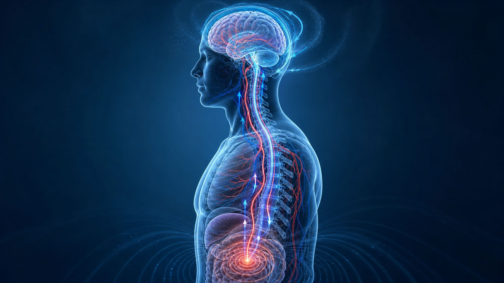
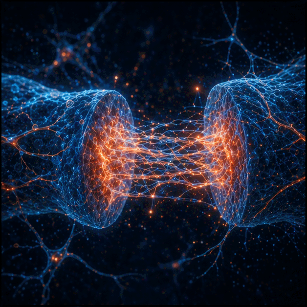
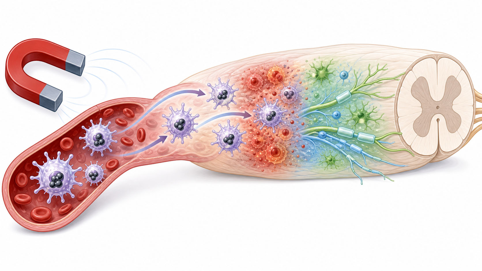
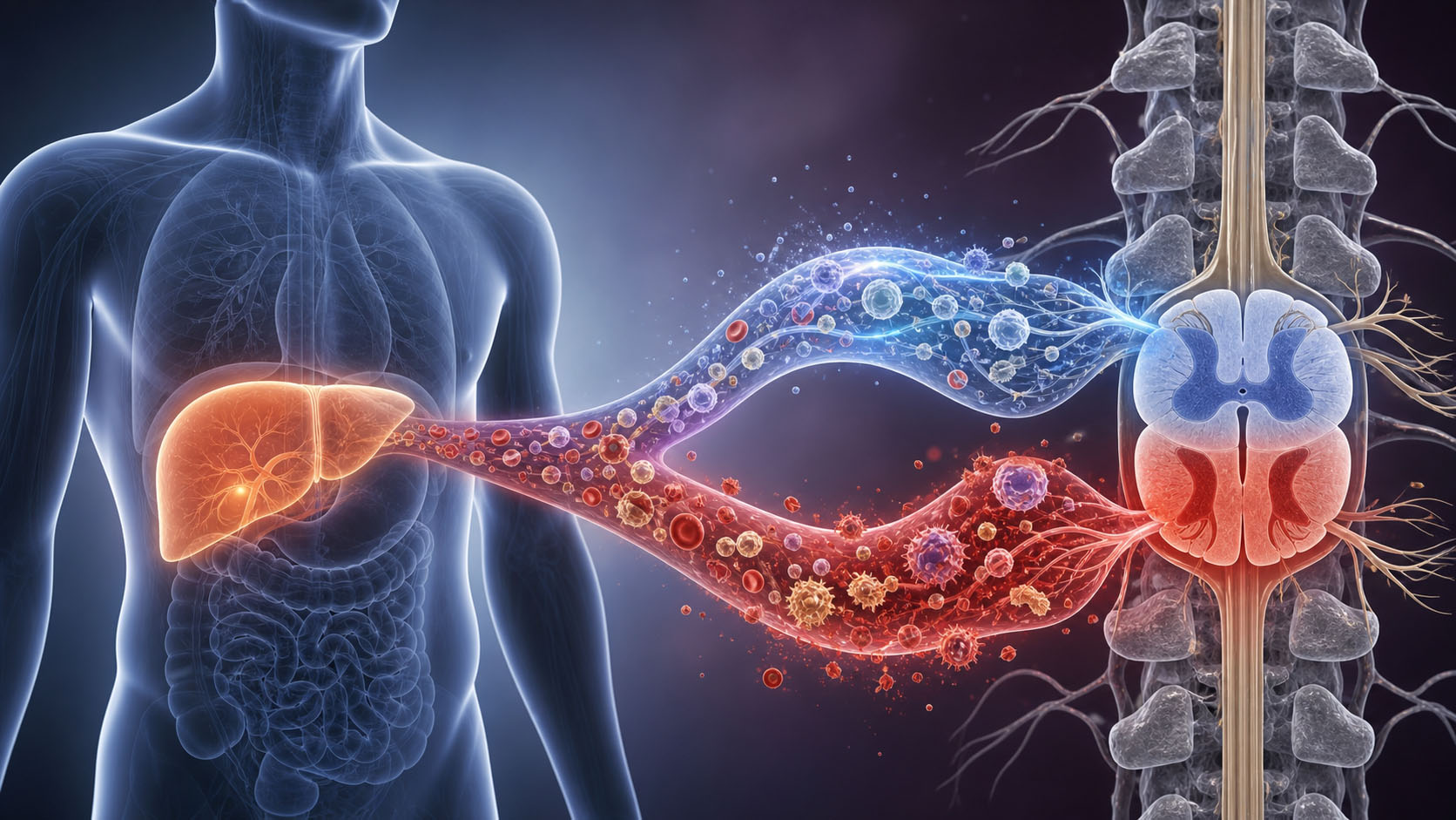
That may sound like a small technical finding, but it could matter. The researchers suggest that abdominal pressure can travel to the brain and spinal canal through a hydraulic-like vascular route, probably involving the vertebral venous plexus. Their modelling also suggests that this brain motion may help move interstitial fluid and cerebrospinal fluid, or CSF, through and out of the brain during wakefulness.

For people with spinal cord injury, this raises an important question: if SCI disrupts abdominal muscle control, autonomic blood pressure regulation, upright movement, and cerebral blood flow, could it also alter this newly described abdomen-brain mechanical system?

The answer is not known yet. But the connection is strong enough to deserve attention.

Author(s):

C. Spencer Garborg, Beatrice Ghitti, Qingguang Zhang, Joseph M. Ricotta, Noah Frank, Sara J. Mueller, Denver I. Greenawalt, Kevin L. Turner, Ravi T. Kedarasetti, Marceline Mostafa, Hyunseok Lee, Francesco Costanzo, and Patrick J. Drew.

Source:

Penn State Neuroscience Institute, Penn State Center for Neural Engineering, The Pennsylvania State University, The University of Auckland, Michigan State University, and collaborating departments. Published in Nature Neuroscience.

SCI context source:

Jill M. Wecht and William A. Bauman, James J. Peters VA Medical Center and Mount Sinai School of Medicine. Their review, "Decentralized cardiovascular autonomic control and cognitive deficits in persons with spinal cord injury", was published in The Journal of Spinal Cord Medicine.

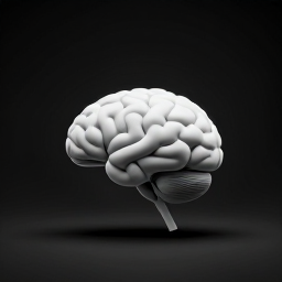
What the brain-motion study found

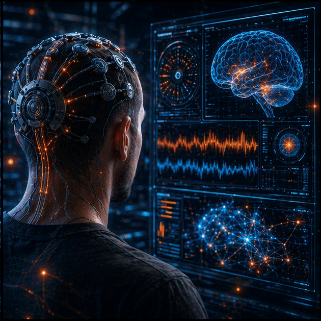
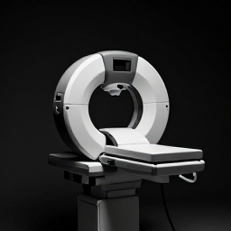
The researchers used high-speed, multiplane two-photon microscopy — a specialist imaging technique that uses laser light to capture movement inside living tissue at very high resolution — to watch the dorsal cortex (the top surface of the brain) move relative to the skull in awake, head-fixed mice. They found that the brain moved mainly rostrally and laterally, meaning forward and sideways.

The movement was tightly linked to locomotion. When the mice moved, the brain moved. But the timing did not match the cardiac cycle or normal respiration. Instead, the key driver appeared to be abdominal muscle contraction.

The team found that abdominal contractions could activate a pressure route between the abdomen and the nervous system. They also showed that applying pressure to the abdomen could induce similar brain motion.

In plain English, the brain may not be as mechanically isolated from the body as we usually imagine. The skull protects it, but pressure changes from the abdomen may still reach the brain through vascular channels connected to the spinal canal.

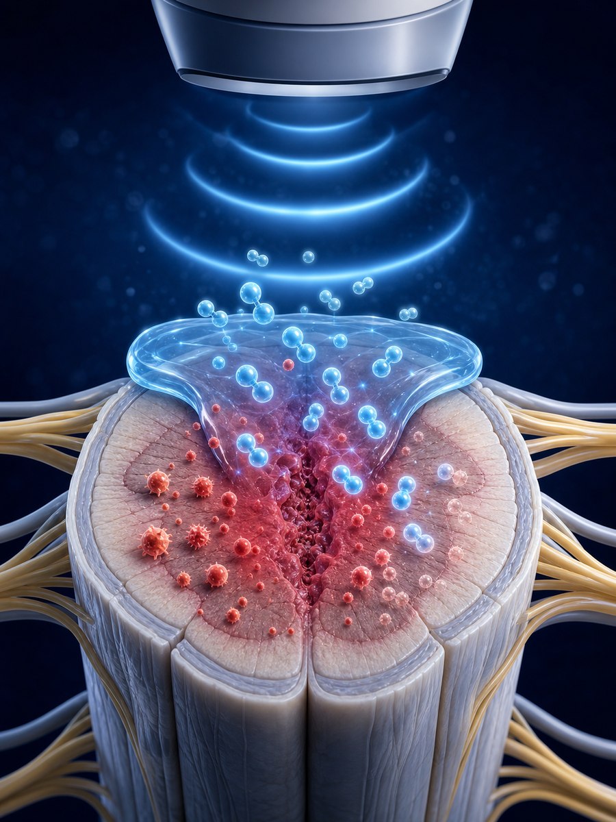
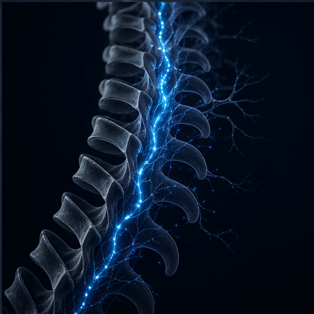
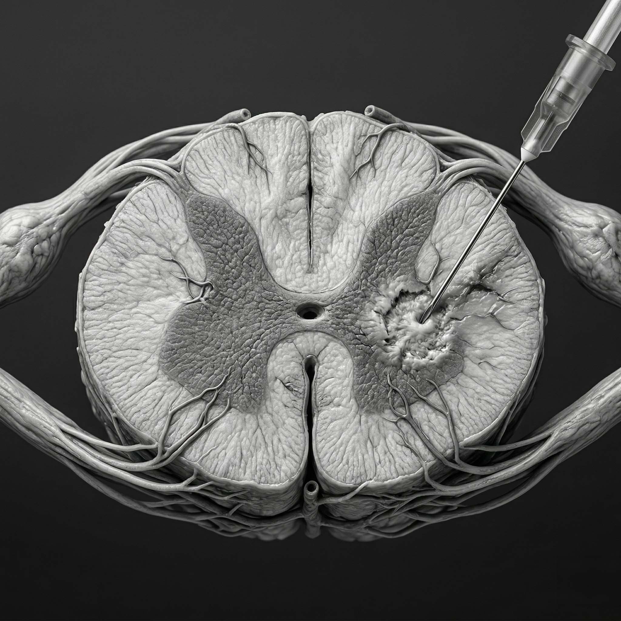
The vertebral venous plexus: a possible pressure pathway

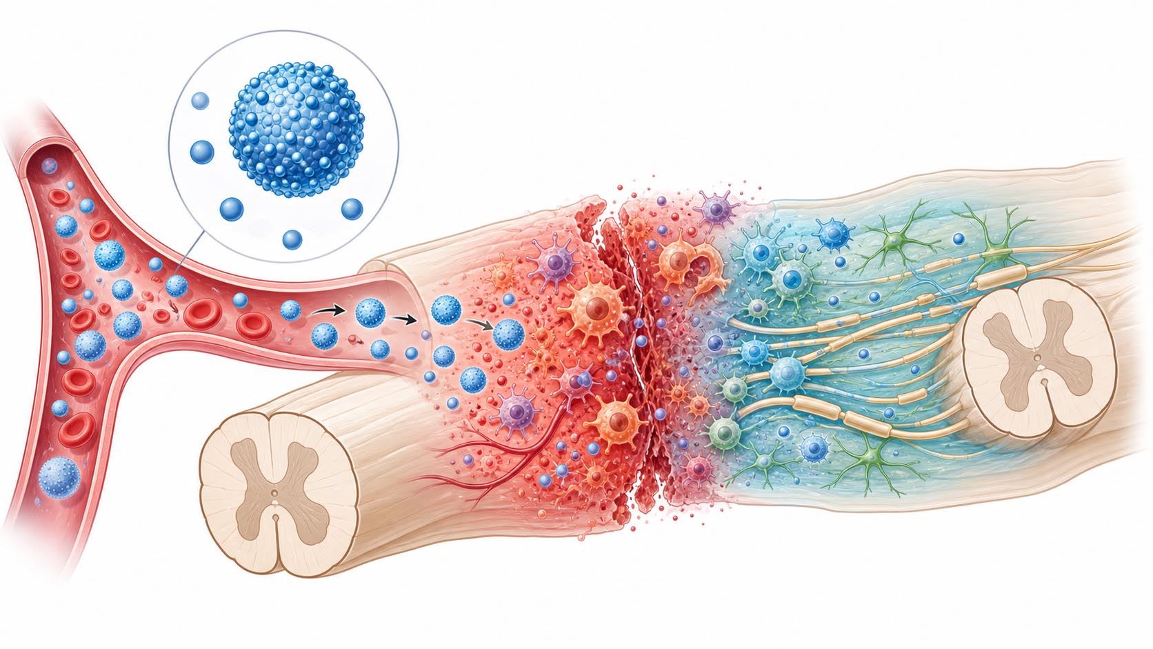
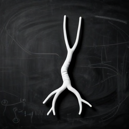
The vertebral venous plexus is a network of veins around the spine. These veins are valveless, meaning pressure can be transmitted through them more freely than through many other blood vessels.

The Nature Neuroscience paper describes this system as a possible hydraulic link between the abdomen and central nervous system. When abdominal pressure rises, that pressure may be communicated to the spinal canal and brain.

A useful analogy is a connected plumbing system. If pressure rises in one chamber, fluid and pressure shifts can affect another chamber connected to it. In this case, the abdomen, spine, and brain may be mechanically linked more than previously appreciated.

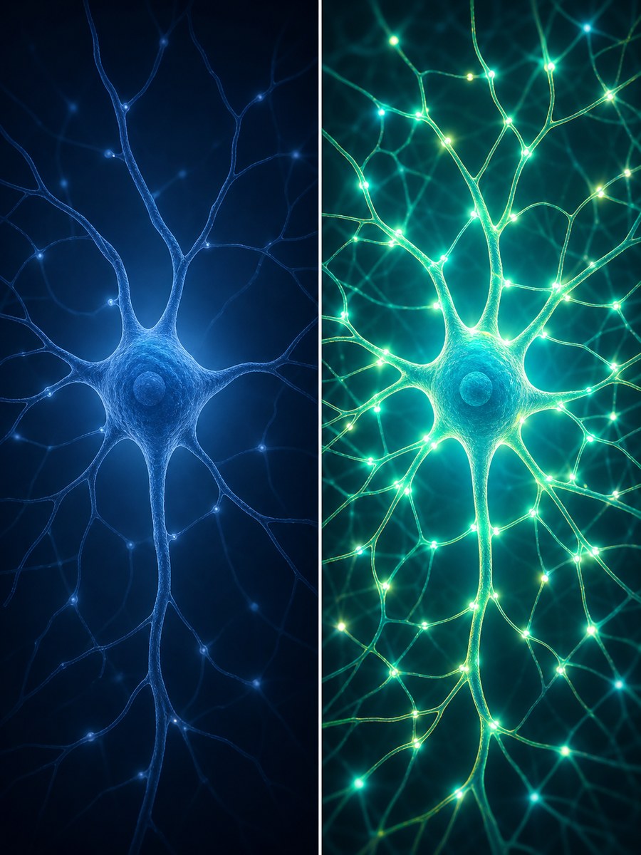
The researchers also suggest that this motion may help drive fluid movement in the brain. That matters because the brain relies on CSF and interstitial fluid movement to help distribute molecules and clear waste. CSF, or cerebrospinal fluid, is the clear liquid that cushions and surrounds the brain and spinal cord. Interstitial fluid is the fluid that fills the tiny spaces between brain cells. Both need to circulate to keep the brain healthy. This is related to the broader field of glymphatic research — the study of the brain's own waste-clearance system, which operates largely during sleep and movement.

Where SCI enters the picture

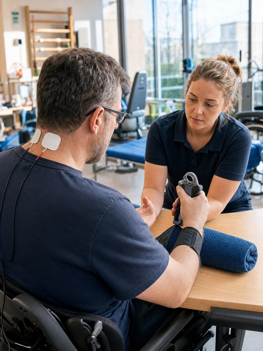
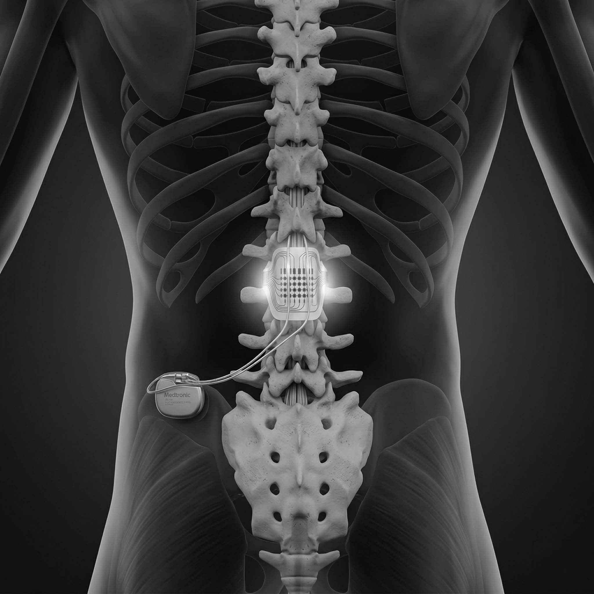
Spinal cord injury often changes far more than movement and sensation. It can disrupt autonomic control: the body's automatic regulation of blood pressure, heart rate, blood vessel tone, sweating, bladder, bowel, and temperature.

The SCI review by Wecht and Bauman describes how cardiovascular autonomic disruption may contribute to cognitive problems after SCI. People with higher injuries may experience low blood pressure, orthostatic hypotension (a drop in blood pressure when sitting up or standing, causing dizziness or faintness), bradycardia (an abnormally slow heart rate), and episodes of autonomic dysreflexia (a potentially dangerous spike in blood pressure triggered by stimulation below the injury level — something many people with cervical or high thoracic injuries will be familiar with). Some people may have reduced resting cerebral blood flow, or a weaker increase in brain blood flow during cognitive tasks.

The review also notes that cognitive deficits after SCI can include problems with memory, attention, processing speed, and executive function. These problems are often blamed on traumatic brain injury or pre-existing factors, but the authors argue that cardiovascular and cerebral vascular dysfunction may also contribute.

This is where the new brain-motion study becomes interesting. It adds another possible layer: body mechanics and pressure-driven brain movement.

A possible new link: pressure, movement, and brain fluid dynamics

In SCI, several things could plausibly affect abdomen-brain mechanical coupling.

First, trunk and abdominal muscle control may be reduced, depending on injury level and completeness. If abdominal muscle activity helps drive brain motion during movement, altered trunk activation could change that mechanical input.

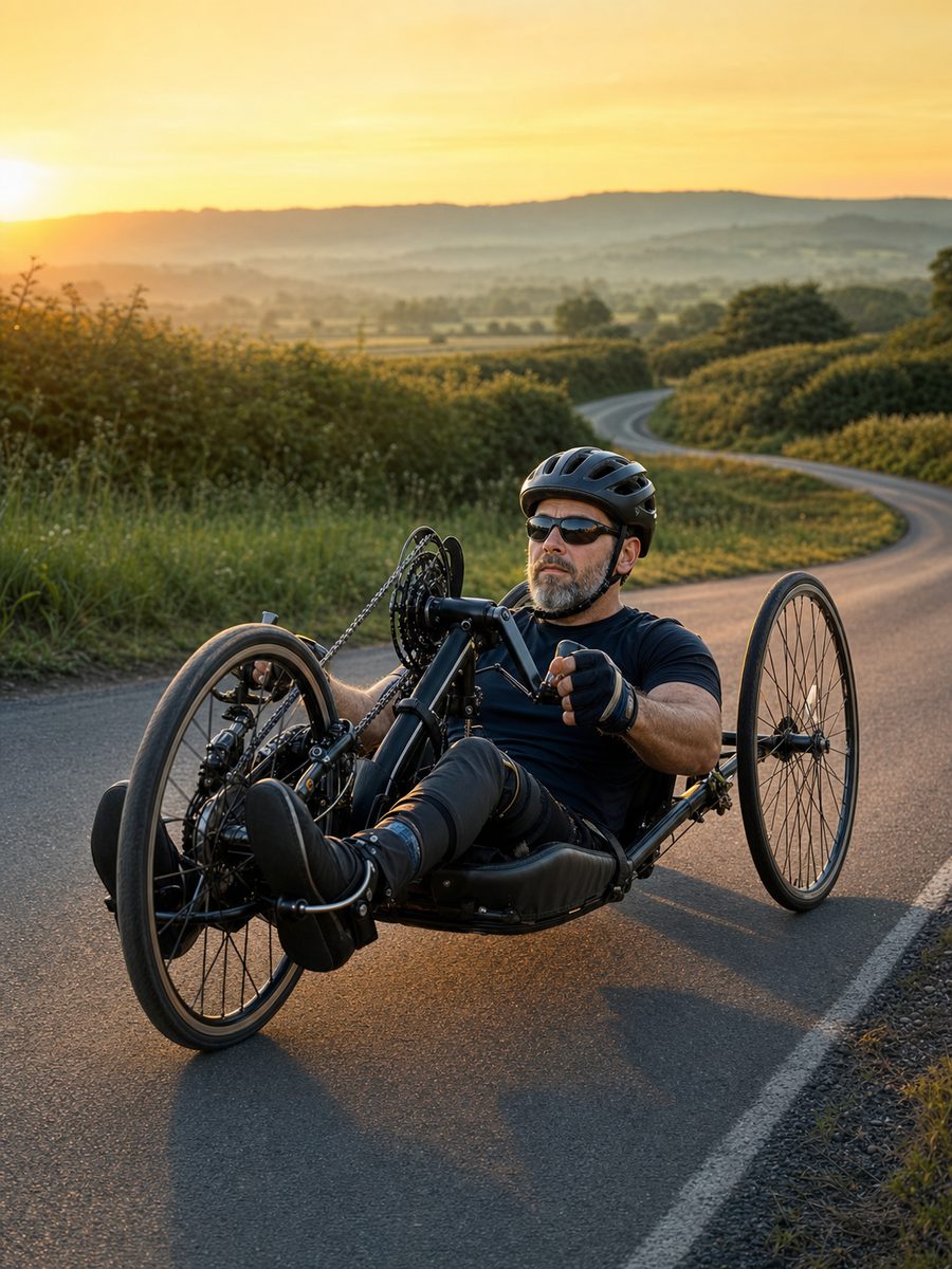
Second, upright movement is often reduced after SCI. Locomotion and body movement were key triggers of brain motion in the mouse study. Less frequent standing, walking, stepping, or trunk-driven movement could mean less of this movement-linked brain fluid activity, though this has not been tested in SCI.

Third, autonomic blood pressure regulation can be unstable. People with SCI may experience low blood pressure, poor orthostatic tolerance, autonomic dysreflexia, or abnormal vascular responses. These could interact with pressure and flow in the spinal and cranial venous systems.

Fourth, bowel and bladder events are already known to be powerful triggers for autonomic dysreflexia in susceptible people. The brain-motion paper discusses abdominal pressure and notes that voiding or defecation can influence pressure states. In SCI, those same pressure events can be medically risky. That makes the connection relevant, but it also means it must be handled carefully.

What this could mean for brain fog after SCI

Many people with SCI describe brain fog, fatigue, light-headedness, poor concentration, or worse thinking when upright, hypotensive, overheated, sleep-deprived, or after autonomic episodes. The established explanation often focuses on blood pressure and cerebral blood flow.

That explanation still matters. If the brain is not getting stable blood flow, thinking can suffer.

The new paper suggests researchers may also need to ask whether pressure-driven brain movement and CSF dynamics are altered when autonomic control, abdominal pressure, movement, posture, and venous flow are changed.

This does not mean brain fog after SCI is caused by reduced brain motion. That would be too strong. But it does suggest a new research question: could altered body-brain mechanics be one contributor to cognitive symptoms in some people with SCI?

Why this should be written carefully

This Nature Neuroscience study was done in mice, not people with SCI. It did not test spinal cord injury, orthostatic hypotension, autonomic dysreflexia, wheelchair users, abdominal binders, bowel care, bladder routines, or cognitive symptoms.

The SCI review is also not claiming that abdominal pressure drives cognitive deficits. It focuses on cardiovascular autonomic control, blood pressure, cerebral blood flow, arterial stiffness, and cognition.

The link between the two papers is therefore a reasoned scientific connection, not a proven clinical fact.

What researchers could test next

A future SCI study could ask whether people with different injury levels show different brain motion, CSF flow, venous pressure dynamics, or cerebral blood flow during posture changes, trunk movement, abdominal compression, respiratory tasks, or safe rehabilitation activities.

Researchers could also examine whether abdominal binders, standing frames, assisted stepping, breathing training, functional electrical stimulation, bowel/bladder states, or autonomic dysreflexia history change brain blood flow or brain fluid movement.

Importantly, any study would need careful safety monitoring. For people at risk of autonomic dysreflexia, abdominal pressure and bowel/bladder triggers are not casual experimental tools.

Reader Q&A

Q: Does this mean abdominal pressure causes brain fog after SCI?

A: No. The study does not prove that. The more careful idea is that abdominal pressure, movement, venous flow, CSF movement, and brain mechanics may be connected. In SCI, where autonomic control and trunk function can be altered, this could become a useful research direction.

Q: Should someone with SCI try abdominal pressure, straining, or Valsalva manoeuvres (bearing down hard — the kind of effort used to equalise ear pressure or during certain exercises) to improve brain fluid flow?

A: No. That could be dangerous, especially for people at risk of autonomic dysreflexia, blood pressure spikes, dizziness, or cardiovascular complications. This story is about research, not a self-treatment.

Q: What type of SCI might this matter most for?

A: It may be most relevant to people with injuries that affect autonomic control, trunk muscles, blood pressure stability, or upright tolerance. Higher-level injuries are especially important because they are more likely to involve hypotension, orthostatic hypotension, and autonomic dysreflexia, but the idea needs direct testing.

The takeaway

This paper gives researchers a new way to think about the body-brain connection. The brain may be mechanically linked to abdominal pressure and movement through vascular pathways around the spine. For SCI, that could matter because injury can disrupt abdominal muscle control, autonomic blood pressure regulation, venous flow, posture, movement, and cerebral blood flow.

The immediate message is not treatment. The message is possibility: brain fog and cognitive fatigue after SCI may deserve investigation not only through blood pressure and brain blood flow, but also through pressure-driven brain and CSF mechanics.

Read full article → (external link)
3

1 MAR 2026

Nerve vs. Tendon Transfers: Major 30-Study Review Finds Nerve Transfers Win for Natural Movement

A massive review of 30 studies has confirmed that while tendon transfers give paralysed hands raw gripping power, nerve transfers provide more natural, fluid movement for everyday tasks.

Deep Dive

When a cervical (neck-level) injury occurs, the hands often lose function entirely. Surgeons use two main 'rewiring' techniques. Nerve Transfers re-route a healthy, redundant nerve from above the injury — like the brachialis nerve in the arm — and connect it to a non-working nerve below. This allows the brain to eventually communicate with the hand again, resulting in natural biomechanics (fluid, complex finger movement). Tendon Transfers are a mechanical fix: a working muscle such as the brachioradialis in the forearm is physically detached and reconnected to a different tendon to create a specific movement like a pinch or grip. The review found that nerve transfers are superior for fine motor control, while tendon transfers are better for generating the raw strength needed for heavy grasping — pointing toward patient-specific surgery planning.

Read full article → (external link)

Latest SCI Developments

Filter by main category

Electrical Stimulation Upper Limb Function 21 MAY 2026

NeuroLife Sleeve Trial Now Recruiting for Hand Function After Cervical SCI

A current ClinicalTrials.gov study is recruiting people with chronic cervical spinal cord injury to test Battelle's NeuroLife EMG-FES Sleeve System, a non-invasive forearm sleeve designed to read attempted muscle signals and help restore grasp through electrical stimulation.

Read More

Deep Dive

For people with cervical spinal cord injury, hand function is not a small detail. It can be the difference between needing help for almost everything and being able to control a little more of your own world.

A current recruiting trial is now testing Battelle's NeuroLife EMG-FES Sleeve System, a wearable forearm sleeve designed to help people with tetraplegia practise grasp and release movements.

The basic idea is simple but powerful. The sleeve tries to detect tiny muscle signals in the forearm when a person attempts to move their hand. These signals are called EMG, short for electromyography. Even when the hand does not visibly move, there may still be weak electrical activity showing that the person is trying.

The second part is FES, or functional electrical stimulation. This uses small electrical pulses through the skin to activate muscles. In the NeuroLife system, the sleeve can both listen for attempted movement and stimulate the muscles needed to help carry out that movement.

Source:

ClinicalTrials.gov study NCT06087445, Battelle's NeuroLife technology page, and previous NeuroLife research involving Ohio State University and Battelle.

What is being tested

The current study is testing the NeuroLife EMG-FES Sleeve System in adults with chronic tetraplegia due to spinal cord injury.

The system is non-invasive. It is worn on the forearm and uses surface electrodes rather than surgery or implanted parts. Trial descriptions say the sleeve can include up to 160 electrodes that record muscle activity and deliver stimulation.

Participants complete a 12-week rehabilitation programme, usually three in-person sessions per week. During these sessions, a therapist works with the participant while they wear the sleeve and practise functional hand and forearm activities.

Where this comes from

This device has a longer backstory than a typical new trial listing.

Battelle and Ohio State University were involved in the earlier NeuroLife neural bypass work, best known through Ian Burkhart. That earlier system used a brain implant, computer decoding, and a forearm stimulation sleeve to help restore voluntary hand movement in a laboratory setting.

That was an important proof of concept, but it was not a practical everyday solution for most people. It required brain surgery and specialist lab equipment.

The newer sleeve approach is different. Instead of reading movement intention directly from the brain, the current NeuroLife EMG-FES system aims to read intention from the forearm muscles themselves. This could make the technology much more practical if future trials show that it is safe and useful.

There was also an earlier completed SCI study, NCT05128994, focused on hand grasp after spinal cord injury. Public final results from that study do not appear to be fully posted yet, but the current recruiting trial suggests the programme has continued into a more structured rehabilitation study.

Why this matters in SCI

A small change in hand function can be a big change in daily life.

Being able to grasp a cup, release an object, use a phone, press a switch, type a few keys, or control a wheelchair joystick can all matter hugely after cervical SCI.

That is why this trial is interesting. It is not promising a cure. It is not claiming to repair the spinal cord overnight. It is asking a more practical question: can a wearable sleeve help people with tetraplegia turn attempted movement into assisted movement?

If the answer is yes, even modest gains could be meaningful.

What this does not prove yet

This remains investigational technology.

Battelle states on its NeuroLife page that the technology is not commercially available and has not been approved or cleared by the FDA for treatment.

The current trial is also small, with a planned enrolment of around 12 participants. It is mainly looking at safety, feasibility, early functional effects, and whether EMG signals could be used as a marker of recovery over time.

In other words, this is promising research, not an available treatment.

Reader Q&A

Is this recruiting now?

Yes. The current ClinicalTrials.gov study is NCT06087445.

Does the sleeve require surgery?

No. This version is non-invasive and uses surface electrodes on the forearm.

Is this the same as the original NeuroLife brain implant system?

No. It comes from the same wider NeuroLife research story, but this version aims to avoid brain surgery by using muscle signals from the forearm instead.

Could it restore normal hand function?

That is not proven. The realistic goal at this stage is to test whether the system is safe, usable, and shows early signs of helping hand and arm function after chronic cervical SCI.

Read full article → (external link)
Electrical Stimulation Spasticity 14 MAY 2026

Dual-Mode Epidural Stimulation Targets Both Spasticity and Movement After SCI

A 2026 preprint reports that lesion-proximal epidural spinal cord stimulation plus physical therapy improved spasticity, pain, sensory scores, and motor measures compared with physical therapy alone in 30 people with traumatic SCI.

Read More

Deep Dive

Spinal cord stimulation is often discussed as a movement technology. But for many people with SCI, movement is not the only problem. Spasticity, pain, stiffness, clonus, and involuntary muscle activity can be just as limiting.

A new preprint study looked at a dual-mode epidural spinal cord stimulation approach designed to do two jobs: calm spasticity and support voluntary movement.

The idea is straightforward but important. One stimulation mode used high-frequency stimulation, around 1.2 kHz, to suppress spasticity. The other used lower-frequency stimulation, 30 to 60 Hz, to help motor facilitation during rehabilitation.

Instead of asking stimulation to do one thing, the researchers used different electrical settings for different nervous-system goals.

Author(s):

Yandong Fan, Mangsuer Nuermaimaiti, Hangfei Guo, Manli Zhu, Lei Ren, Yirizhati Aili, Mamutijiang Muertizha, Aihemaiti Aierken, and Kun Luo.

Source:

First Affiliated Hospital of Xinjiang Medical University, Xinjiang Medical University, and Second Affiliated Hospital of Xinjiang Medical University. Posted as a Research Square preprint.

What was tested

The study included 30 people with traumatic spinal cord injury. Participants were randomised to either epidural spinal cord stimulation plus physical therapy, or physical therapy alone.

The stimulation group received a 16-contact epidural electrode implanted close to the injury, one to two spinal segments above the lesion. The authors describe this as lesion-proximal placement.

This differs from many earlier stimulation approaches that mainly targeted intact lumbar circuits below the injury. Here, the researchers aimed to modulate the region nearer the damaged segment.

The stimulation was adjusted during surgery and then reviewed monthly.

Why two modes?

Spasticity and movement facilitation can pull in opposite directions.

Low-frequency stimulation may help activate motor circuits and support voluntary movement. But if someone has severe spasticity, making the nervous system more excitable can sometimes worsen stiffness, spasms, clonus, or unwanted co-contraction.

High-frequency stimulation can be used differently: to dampen overactive reflex pathways.

A simple analogy is driving a car with both a throttle and a brake. If the brake is stuck, pressing the throttle harder does not give smooth movement. This dual-mode approach tries to release the brake first, then support useful movement.

What the study reported

At 30 days, the stimulation plus physical therapy group showed better outcomes than physical therapy alone in several areas:

- Sensory improvement: 93.3% versus 60.0%. - Spasticity control: 73.3% versus 26.7%. - Pain reduction: 60.0% versus 20.0%.

At six months, the stimulation group showed sustained gains. ASIA sensory scores increased, motor scores improved, Modified Ashworth Scale scores fell, and pain scores improved.

The authors report that 80% of the stimulation group achieved clinically meaningful functional improvement, including recovery involving ankle dorsiflexors and gait-related muscles.

For readers, ankle dorsiflexion matters because it helps lift the front of the foot during stepping. Even partial improvement in that movement can be functionally meaningful.

Why this is relevant to spasticity

Spasticity is not just tight muscles.

It can include exaggerated reflexes, increased tone, clonus, involuntary spasms, and muscles firing against each other during attempted movement. That can make transfers, sitting, sleep, hygiene, bladder and bowel routines, and exercise harder.

Many drug treatments can help but may cause side effects such as weakness, tiredness, low tone, dizziness, or withdrawal problems. For some people, spasticity remains difficult to control.

This study is interesting because it treats spasticity as a central part of recovery, not as a side issue.

What this does not prove yet

This is a preprint, meaning it has not completed final peer review.

The study is also relatively small, with 30 participants. The results are promising, but they need independent confirmation, clearer reporting of safety outcomes, and longer-term follow-up.

Epidural stimulation also requires surgery. That means risks, screening, specialist implantation, programming, and rehabilitation support. It is not comparable to a simple external device.

The paper also sits in an interesting context. Other recent stimulation research warns that waveform, frequency, electrode placement, and target fibres all matter. High-frequency stimulation may help with some goals, such as suppressing spasticity, but it should not be assumed to be automatically useful for every recovery target.

Why this matters in SCI

The most useful part of this study is the treatment logic: one stimulation setting may not fit every symptom.

People with SCI may need stimulation programmes that change depending on the goal: relaxing spasticity, supporting stepping, assisting voluntary movement, reducing pain, or stabilising autonomic function.

If future trials confirm these findings, dual-mode stimulation could help move SCI neuromodulation toward personalised programming rather than fixed, one-size-fits-all settings.

Reader Q&A

Could this help someone with severe spasticity?

Possibly in the future, but this is still a preprint and involves implanted epidural stimulation. It should be viewed as promising clinical research, not an available general treatment.

Does high-frequency stimulation restore movement?

Not by itself. In this study, high-frequency stimulation was used mainly to suppress spasticity, while lower-frequency stimulation was used to support motor facilitation.

Why place electrodes near the injury?

The authors argue that placing electrodes close to the lesion may help modulate the injured segment itself, rather than only stimulating intact circuits below the injury. This needs further validation.

Read full article → (external link)
Stem Cells Research Models 14 MAY 2026

Human Lumbar Spinal Cord Organoids Could Give SCI Researchers a Better Lab Model

Researchers in Australia developed human stem-cell-derived spinal cord organoids patterned toward a lumbar identity, creating a more realistic 3D model for studying lower spinal cord biology, motor neurons, neurodegenerative disease, and spinal cord injury.

Read More

Deep Dive

Some spinal cord injury breakthroughs are not treatments. They are better tools.

A new Scientific Reports paper describes a method for growing human spinal cord organoids from induced pluripotent stem cells, or iPSCs. These organoids were guided toward a lumbar spinal cord identity, meaning they resemble the lower spinal cord region more than previous models that often lean toward cervical or thoracic identity.

That matters because the lumbar spinal cord contains motor circuits involved in the legs. For SCI, ALS, spinal muscular atrophy, and other motor neuron conditions, researchers need better ways to study human spinal cord tissue in the lab.

Author(s):

Li Jun Loh, Pratibha Panwar, Shila Ghazanfar, Kwaku Dad Abu-Bonsrah, Forough Habibollahi, Joel Mason, Brett J. Kagan, Bradley J. Turner, and Samantha K. Barton.

Source:

Florey Institute of Neuroscience and Mental Health, University of Melbourne, University of Sydney, Charles Perkins Centre, Sydney Precision Data Science Centre, and Cortical Labs. Published in Scientific Reports.

What is an organoid?

An organoid is a tiny 3D tissue model grown in the lab.

It is not a full organ. It does not think, feel, or work like a complete spinal cord. But it can contain several cell types arranged in a more natural 3D structure than flat cells grown on a dish.

A useful analogy is the difference between studying a city from a list of buildings versus looking at a small 3D neighbourhood model. A flat cell culture can tell researchers a lot about one cell type. An organoid can show more of the relationships between cells.

Why lumbar identity matters

The spinal cord is not the same from top to bottom.

Cervical regions help control the arms and hands. Thoracic regions are involved in trunk and autonomic functions. Lumbar regions are important for lower limb circuits.

Many previous spinal cord organoid systems have been more cervical or thoracic in character. This new protocol aimed to push the organoids toward a caudal, or lower-spinal, fate. The researchers used developmental signals to increase HOXC10, a marker linked to lumbar identity, while reducing more anterior spinal cord identity.

In plain English, they tried to tell the stem cells: "do not become generic nervous-system tissue; become more like lower spinal cord tissue."

How the organoids were made

The team started with human iPSCs. These are adult-derived cells reprogrammed into a stem-cell-like state, meaning they can be guided to become many different cell types.

The researchers then used a sequence of chemical signals to mimic development. These included signals that help establish nervous-system fate, caudal spinal identity, ventral patterning, and motor neuron development.

Over time, the cells formed 3D spheres containing spinal cord-like cell populations.

What the researchers found

The organoids contained an enriched neuronal population along with several glial cell types.

Glia are support cells of the nervous system. They are not just background helpers. Astrocytes, oligodendrocyte-lineage cells, and other glial populations influence survival, inflammation, myelin, metabolism, and how neurons function.

That is important because SCI is not just a neuron problem. Injury affects neurons, glia, myelin, blood vessels, immune responses, and the tissue environment.

Single-cell RNA sequencing showed that the organoids contained diverse cell populations and better recapitulated ventral spinal cord identity than standard 2D motor neuron cultures.

The organoids also showed spontaneous electrical activity, suggesting that the cells were forming functional neural networks.

Why this could help SCI research

Animal models remain essential in SCI research, but they are not human.

Human spinal cord tissue is difficult to study directly, especially during early disease or injury processes. Organoids can help bridge that gap. They allow researchers to study human cells in a controlled environment, test drugs, explore disease mechanisms, and compare patient-specific cell lines.

For SCI, lumbar organoids could be useful for studying:

- How human motor neurons mature and connect. - How glial cells affect motor neuron survival. - How injury-like stress affects spinal cord cell networks. - How candidate drugs influence human spinal cord cells. - Why some motor neuron populations are more vulnerable than others.

This does not replace animal studies or clinical trials. It gives researchers another tool before moving into those harder and more expensive stages.

What this does not mean yet

These organoids are not transplant-ready tissue.

They are a model system. They do not contain the full structure of a spinal cord, long-distance body connections, blood supply, immune system, pain pathways, movement circuits, or the mechanical environment of an injured spine.

They also model development more than adult injury. Researchers would still need to adapt and validate them for specific SCI questions.

Why this matters in SCI

SCI research needs better human models. Too many potential therapies look promising in animals or simple cell cultures and then fail later.

A more realistic human lumbar spinal cord organoid could help screen ideas earlier and more accurately. It may also support personalised research in the future, where organoids made from a person's own cells are used to understand disease risk or test treatment responses.

This is not a cure story. It is a better-map story.

Reader Q&A

Could these organoids be implanted into someone with SCI?

No. This study is about creating a lab model, not a transplant therapy. Implantation would require a completely different level of safety and functional testing.

Why is this relevant if it is not a treatment?

Better lab models help researchers choose better treatments to test. They can reduce false starts and make it easier to study human spinal cord biology.

Why focus on the lumbar spinal cord?

The lumbar region contains circuits important for lower limb function. A lumbar-like model may be more relevant for studying leg-related motor neurons and lower spinal cord disease mechanisms.

Read full article → (external link)