Insights into all the latest

Research in the News

Tracking the pulse of the latest spinal cord injury research news — from lab breakthroughs to clinical milestones.

Author's Current Top 3 to Read

1

31 JUL 2026

Breathing After High Cervical SCI Without a Ventilator: The Surgery More People Should Know Exists
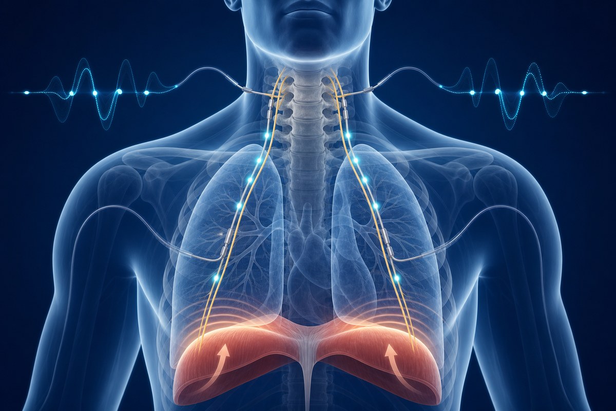
When two-year-old Maverick sustained a high cervical spinal cord injury in a crash, his parents were told he would never breathe without a machine. In January 2026, surgeons at Oklahoma Children's made him the youngest person in the world to undergo phrenic nerve stimulation — a procedure that bypasses the injured cord to restore breathing. Here's what it is, who it can help, and why it isn't more widely known.

Deep Dive

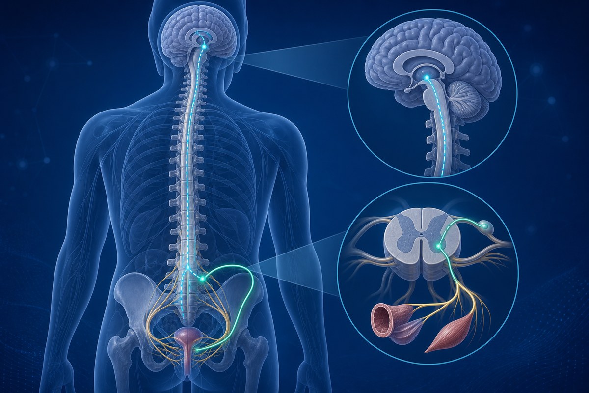
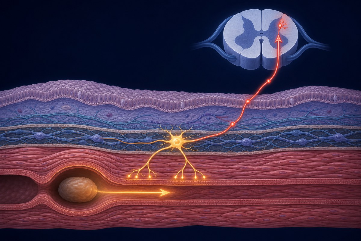
Most people with a high cervical spinal cord injury spend the rest of their lives on a ventilator. Not because their lungs have failed. Not because their diaphragm has stopped working. But because the signal the brain sends to start breathing — travelling down through the spinal cord to the phrenic nerve, the nerve that controls the diaphragm — can no longer get through.

That signal pathway is broken at the cord. But the phrenic nerve itself, in most high cervical injuries, is completely intact. And that is the opening that phrenic nerve stimulation — also called diaphragmatic pacing — exploits.

On 30 July 2026, Oklahoma Children's OU Health published the story of Maverick, a Missouri toddler who suffered a catastrophic cervical SCI in a road crash at 18 months old. He became the youngest person in the world to have phrenic nerve stimulators implanted following a spinal cord injury — at just over two years old, in January 2026. He is now breathing with his own diaphragm, electrically triggered, without a ventilator for stretches of time and progressing further.

His family says they wish more people knew this procedure existed. They're right to say so.

Why breathing fails after high cervical SCI

The phrenic nerves originate in the cervical spinal cord at the C3, C4, and C5 levels — roughly mid-neck. They travel down through the chest and connect to each side of the diaphragm, the dome-shaped muscle that does most of the work of breathing. When the brain signals a breath, the message travels down the cord, exits at C3–C5, and drives the diaphragm to contract and pull air into the lungs.

A spinal cord injury above C3 severs this chain at the cord. The phrenic nerves below the injury remain intact. The diaphragm remains intact. There is simply no signal reaching either of them.

This is where the ventilator steps in: it pushes air into the lungs mechanically, doing the work the diaphragm can no longer do. It is life-saving. But it has real downsides — which is exactly why phrenic nerve stimulation exists.

What phrenic nerve stimulation is

The procedure places small electrodes surgically around or near the phrenic nerve (or at the nerve's entry points into the diaphragm, depending on approach). These electrodes connect to a receiver implanted under the skin. An external control unit transmits electrical pulses through the skin to the receiver, which passes them on to the electrodes, which cause the phrenic nerve to fire — and the diaphragm to contract. A breath follows.

The timing and intensity of those pulses are adjusted to produce a normal breathing rhythm. With conditioning over weeks to months — the diaphragm is a muscle, and needs to be rebuilt after disuse — many patients go from a few hours off the ventilator each day to spending most or all of the day breathing with the pacer. Some transition off the ventilator entirely.

There are two main device types. The conventional approach uses an electrode on the phrenic nerve connected to a subcutaneous receiver and an external radiofrequency transmitter worn on the skin. The Diaphragmatic Pacing System (DPS) places four electrodes directly into the diaphragm at the nerve insertion points, connected to an external pulse generator through a socket at the skin surface. Each approach has clinical uses; the DPS in particular allows stimulation even when the phrenic nerve higher up is partially compromised.

Who qualifies

The key requirement is that the phrenic nerve itself must be intact and functional. Before surgery, phrenic nerve conduction studies — electrical tests of nerve function — confirm whether the nerve can carry a signal.

Patients with SCI above C3 are the clearest candidates: the injury is above where the phrenic roots exit the cord, so the nerve below is undamaged and can be stimulated at the neck, chest, or diaphragm.

Mid-cervical injuries (at C3–C5 itself) are more complex, since the injury may directly involve the nerve roots. In these cases, direct diaphragmatic pacing through the DPS system can sometimes still work by stimulating the nerve at the diaphragm rather than higher up.

Beyond SCI, the procedure is used in congenital central alveolar hypoventilation syndrome (where the brain's breathing control fails), brainstem tumours, Arnold-Chiari malformations, and certain neuromuscular conditions.

How the surgery is done

Surgeons can reach the phrenic nerve three ways. The cervical approach accesses the nerve directly in the neck — the most direct route, typically used for high SCI. The thoracic approach uses keyhole video-assisted chest surgery (VATS) to reach the nerve as it runs alongside the heart. The diaphragmatic approach is laparoscopic — small incisions in the abdomen — and places electrodes where the nerve meets the diaphragm. This is the route used for the DPS device and is available even when the nerve higher up is unreliable.

Both sides are usually done two weeks apart to allow recovery between procedures.

Why it beats a ventilator for most patients

Ventilators save lives, and many people with high SCI live full lives on them. But the practical burden is real: a ventilator requires a tracheostomy (a permanent opening in the windpipe), carries ongoing infection risk, needs an uninterrupted power supply, and is a disconnection risk. Speaking is harder. Taste diminishes over time. Mobility is constrained.

Phrenic nerve stimulators are a closed system once implanted. Most patients keep their tracheostomy initially as a safety backup, but the breathing itself comes through the pacer rather than the machine. Speech follows the natural rhythm of the pacer. Taste is preserved. The device is small and unobtrusive. Because the diaphragm contracts naturally — even if electrically triggered — breathing mechanics are closer to normal, which is better for long-term lung health.

In February 2026, NICE in the UK issued guidance (HTG727) formally endorsing phrenic nerve pacing for ventilator-dependent patients with high cervical SCI — a signal that clinical consensus around the technology is consolidating.

Why it isn't more widely known

Phrenic nerve stimulation has existed since the 1970s — Christopher Reeve used a diaphragmatic pacer in his later years. The technology has improved significantly: devices are smaller, laparoscopic implantation is less invasive than early open surgery, and FDA approval has made the DPS accessible across the US.

But it remains a specialist procedure, concentrated at a small number of centres with specific experience. Awareness among patients, families, and even some clinicians is still limited — which means people who might benefit are sometimes not told it exists. Maverick's case, as the youngest patient in the world to undergo the procedure after SCI, brings it to a wider audience at a moment when guidance and availability are both moving in the right direction.

Reader Q&A

Does everyone with a high cervical SCI qualify?

Not automatically — phrenic nerve function has to be confirmed first through nerve conduction tests. Most people with injuries above C3 do qualify, since the phrenic nerve below the injury is typically intact. People with injuries at C3–C5 need careful assessment, but some still qualify via the diaphragmatic approach. A specialist centre with experience in the procedure is the right place to be evaluated.

Can it replace the ventilator completely?

For many patients, yes, at least for daytime use. Some manage without the ventilator at all. The process involves a conditioning phase — the diaphragm has usually weakened from disuse and needs time to rebuild — which takes weeks to months. Others use the pacer during the day and non-invasive ventilation at night. The combination depends on the individual. Tracheostomies are often kept in place initially as backup, and removed later once breathing is stable.

What age can it be used from?

Maverick's surgery extends the known lower age limit to just over two years old. The Christopher & Dana Reeve Foundation notes that paediatric use of diaphragmatic pacing has actually driven adoption faster than in adults, partly because children's developing nervous systems are more adaptable. Surgeons experienced in paediatric implantation are needed for young patients — but the procedure is not age-restricted in principle.

Is it available in the UK?

Yes, at specialist centres, and with growing clinical support. The February 2026 NICE guidance (HTG727) formalises it as a recommended option for ventilator-dependent high cervical SCI patients, which should strengthen access through NHS pathways. Referral to a specialist spinal or respiratory centre is the starting point.
Read full article → (external link)
2

15 JUL 2026

How to train your breathing after SCI: intensity beats everything else

A study of 81 people with SCI at Switzerland's leading rehabilitation centre found that training intensity and duration predict respiratory improvement far more than injury level or personal characteristics.

Deep Dive

If you have a spinal cord injury — particularly at a cervical or high thoracic level — the muscles that control your breathing are likely affected. Most people with SCI know this in a general way: reduced lung capacity, difficulty clearing the chest after illness, or shortness of breath with effort. What's been less clear is which training approach actually improves this, and whether the severity of your injury limits what's possible.

New research from the Swiss Paraplegic Centre in Nottwil — one of Europe's leading SCI rehabilitation hospitals — gives a more concrete answer than we've had before. The headline finding is both simple and actionable: train hard, train often, and keep going for at least three months. Almost everything else is secondary.

What the study did

Researchers analysed data from 81 people with SCI who completed respiratory muscle training programmes at the Swiss Paraplegic Centre. Participants spanned a range of injury levels and severities — both complete and incomplete injuries — as well as different ages, body types, and baseline fitness levels. This mix was deliberate: the researchers wanted to understand which factors predicted better respiratory outcomes at the end of training.

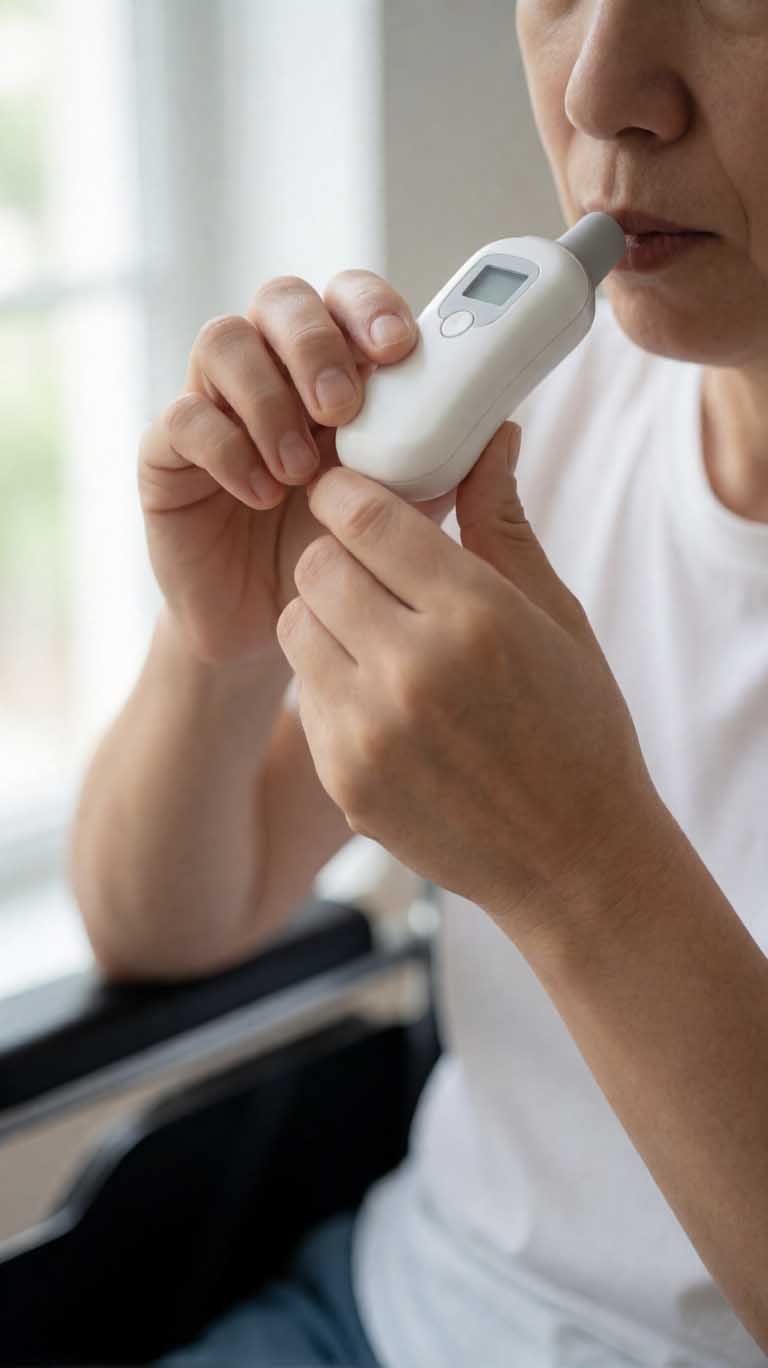
The training used inspiratory muscle training (IMT) devices — small handheld tools that add resistance to your breathing, similar in principle to a breathing weight. Participants trained at varying intensities and for varying durations as part of their rehabilitation plans.

What they found

When the researchers modelled which factors predicted improvement in respiratory muscle strength, training intensity and programme duration emerged as the dominant predictors — by a significant margin. Personal characteristics like age, sex, and body weight had comparatively little predictive power. Injury characteristics — level, AIS classification (complete vs incomplete), and time since injury — also mattered far less than the training variables themselves.

The practical implication is striking: someone with a complete C4 injury who trains consistently at high intensity for four months is likely to see more improvement than someone with an incomplete C6 injury who trains at moderate intensity for six weeks. The injury doesn't cap your gains as much as the training approach defines them.

The researchers' specific recommendations: train at the highest intensity you can sustain, three to five times per week, for a minimum of three months.

Why respiratory muscle strength matters so much

Pneumonia is the leading cause of death in people with AIS A (complete motor and sensory loss) and AIS B (complete motor loss, some sensory preserved) spinal cord injuries. The primary reason is that impaired respiratory muscles make it harder to cough forcefully and clear the airways of secretions — a problem that becomes critical during a chest infection.

Stronger respiratory muscles translate directly into a more effective cough, better ability to manage respiratory illness at home, and — critically — a reduced risk of pneumonia requiring hospitalisation or mechanical ventilation. The stakes are high enough that this is one area of rehabilitation where the evidence really matters.

Why this study is worth paying attention to

The Swiss Paraplegic Centre treats around 300 new SCI patients a year and carries one of the largest longitudinal SCI rehabilitation datasets in the world. Research from this centre tends to be grounded in clinical reality rather than tightly controlled experimental conditions — these were real patients doing real rehabilitation, not a highly selected research cohort. That makes the findings more directly applicable to practice.

The study also addresses a question that's been genuinely uncertain: does it matter who you are, or does it matter what you do? The answer, clearly, is the latter. That's useful information for patients deciding whether to invest in a training programme, and for clinicians deciding how to structure one.

Reader Q&A

Should I be doing respiratory muscle training? If you have a cervical or upper thoracic SCI and you ever notice difficulty clearing your chest, shortness of breath, or recurring respiratory illness, then yes — it's worth discussing with your physiotherapist or rehabilitation doctor. The evidence is now reasonably strong that structured training helps, and the risk is low.

What kind of device is used? Inspiratory muscle training (IMT) devices are small handheld tools you breathe through against adjustable resistance. They're available without prescription and relatively inexpensive. Your physiotherapist can advise on the right resistance level and technique for your situation.

Does this apply to incomplete injuries too? Yes. The study included both complete and incomplete injuries, and the key finding — that training intensity matters more than injury characteristics — applied across the range.

What if I've had my SCI for years? Is it too late to benefit? Time since injury was not a strong predictor of training response in this study, which suggests it's never too late to benefit. That said, starting respiratory training earlier in rehabilitation tends to establish better habits and baselines — so earlier is generally better, but later is still worth doing.

Read full article → (external link)
3

15 JUL 2026

China performs first commercial brain-computer interface surgery for spinal cord injury

On July 13, a 35-year-old man with cervical SCI became the first person in the world to receive a commercially approved implanted brain-computer interface, during a six-hour surgery at Huashan Hospital in Shanghai.

Deep Dive

Last Sunday, a surgical team at Huashan Hospital — one of China's most prominent neurosurgical centres, affiliated with Fudan University in Shanghai — spent six hours implanting a small array of electrodes into the brain of a 35-year-old man who had lost voluntary use of his hands following a cervical spinal cord injury.

This wasn't an experimental procedure in the usual sense. The device they used — the NEO system, developed by Chinese company Neuracle Technology — had just received commercial approval from China's National Medical Products Administration (NMPA), the country's equivalent of the FDA. This surgery was the first performed under that approval.

If that sounds significant, it is. Until last week, no implantable brain-computer interface had ever been granted commercial approval anywhere in the world. Every previous BCI implanted in a person with paralysis — including those in US Neuralink trials — was done under research exemptions, not as a licensed product a doctor could simply prescribe. We covered the regulatory milestone when it was announced earlier this week; Sunday's surgery is the next chapter.

What the device does

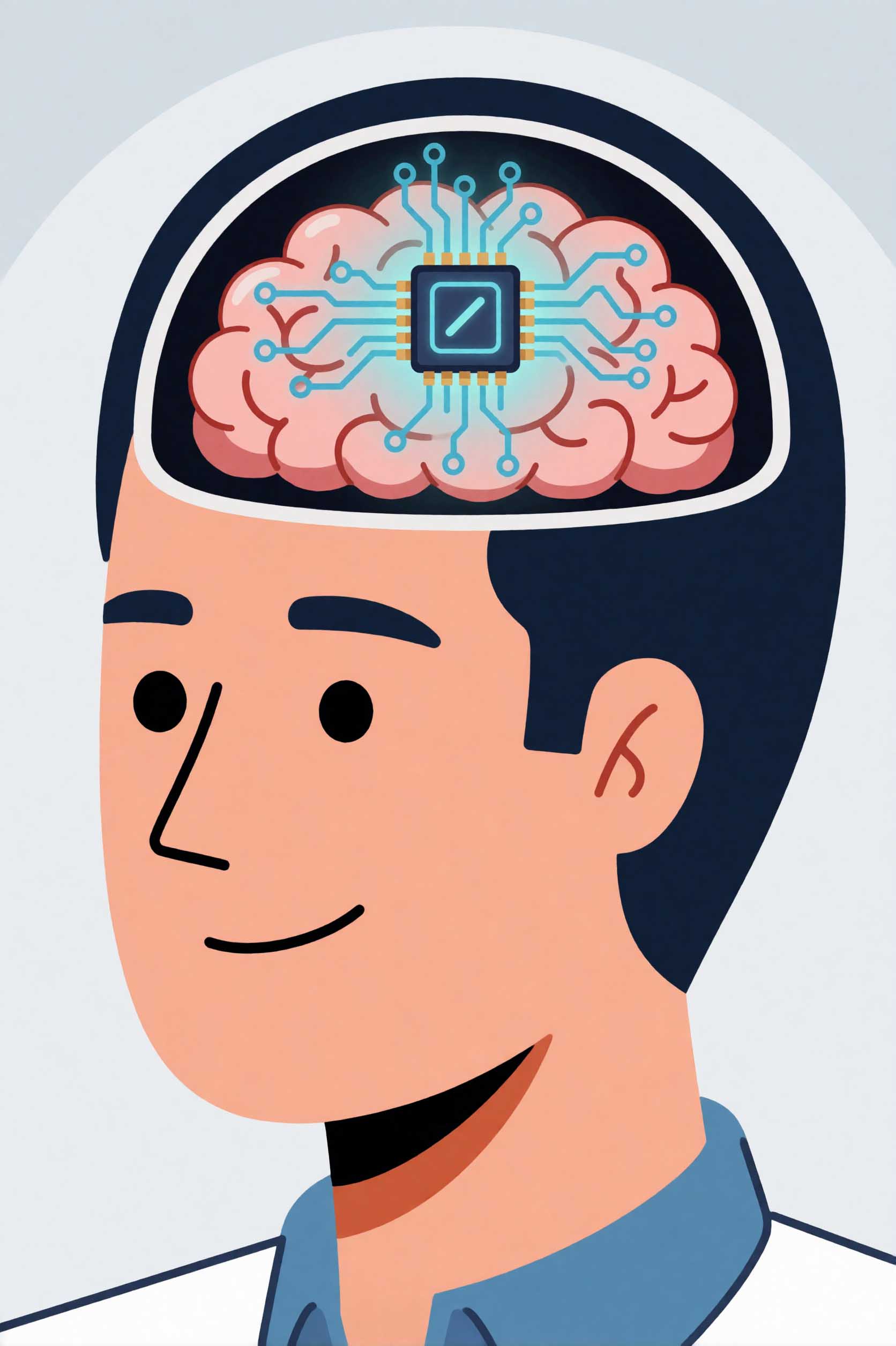
The NEO system is designed for people with cervical SCI — specifically those with injuries between C3 and C7 — who have lost voluntary control of their hands and fingers. The device consists of electrodes implanted into the motor cortex, the part of the brain that controls voluntary movement. It reads the electrical signals the brain generates when the person *thinks* about moving their hand, and translates those signals into commands that can stimulate muscles or control external devices.
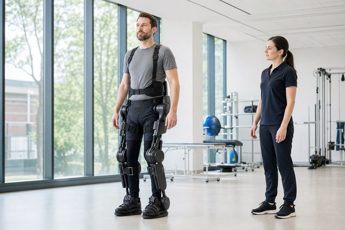
The goal is what researchers call "thought-controlled muscle rehabilitation" — restoring functional hand use by creating a new pathway between brain and muscle that bypasses the damaged cord.

Following the surgery, the patient will need to commit to six hours of rehabilitation per day to train the system and rebuild functional connections. That's intensive, and it underscores that this isn't a passive implant — it requires significant sustained effort from the person receiving it.
Why China, and why now

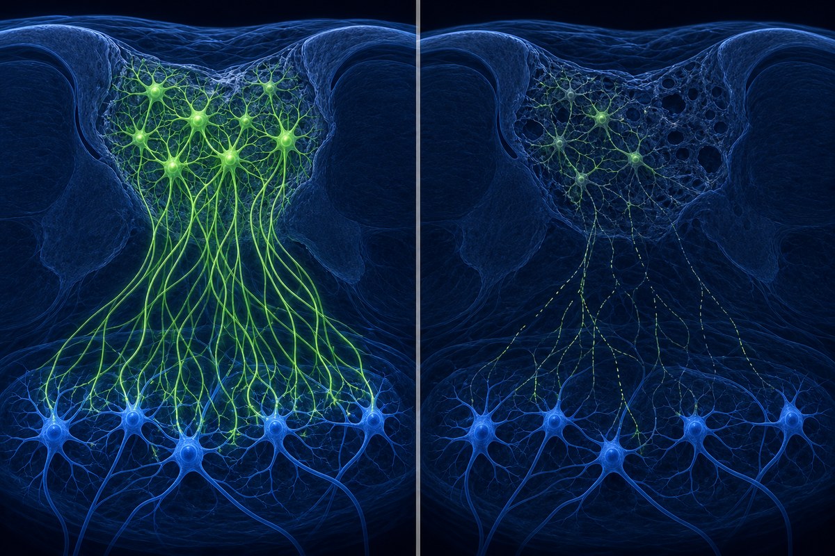
China has an estimated 3.7 million people living with spinal cord injuries — one of the largest SCI populations in the world. Neuracle Technology has been developing the NEO system for several years, with clinical trials demonstrating safety and some functional benefit sufficient for the NMPA to grant commercial approval.
The specific evidence package used to support the approval hasn't been published in full, but the fact that a sovereign national regulator — with its own evidence standards — granted commercial status to an implantable BCI is notable. No other country has done this yet.

What this doesn't mean yet

It would be easy to read "commercially approved BCI" and assume this technology is now broadly available. It isn't. The NEO system is approved in China. It has not been reviewed by the FDA, the European Medicines Agency, or the MHRA. The approval pathway in China, while rigorous, differs from Western jurisdictions, and each regulator will require its own evidence review.

Access will also depend on cost, surgical infrastructure, and — critically — post-surgical rehabilitation resources. Six hours of therapy daily is a serious commitment, one that requires substantial clinical support to sustain.

None of this diminishes what happened on Sunday. For the first time in history, an implantable BCI for paralysis has moved from experimental to commercial, and the first patient has received it. The regulatory door has been opened. The scientific and commercial pressure to bring equivalent devices through the FDA and European processes will only grow.

Reader Q&A

Could someone with SCI get this surgery right now? Not outside China, and even within China, access will depend on cost, clinical eligibility, and availability of surgical centres experienced with the procedure. This is a brand-new commercial product — the first surgery was performed last Sunday.

Is the NEO system the same as Neuralink? No. Neuralink is a US company with its own design still in clinical trials. Neuracle Technology is a separate Chinese company whose device has now received commercial approval — a stage Neuralink has not yet reached.

Does this mean there's a cure for hand paralysis after SCI? No. The NEO system creates a new communication pathway between brain and muscles, but it doesn't repair the spinal cord. It's an assistive technology that, with intensive rehabilitation, may allow some people to regain functional hand use. The extent of recovery will vary by individual.

When might something like this be available in the UK, US, or Europe? Genuinely unknown. It would require separate regulatory submissions, clinical trials acceptable to the FDA or MHRA, and likely several more years of development. The China approval is a significant milestone, but it doesn't shorten any other regulator's pathway.
Read full article → (external link)

Latest SCI Developments

Filter by main category

Research Epidemiology 28 SEPT 2026

In 366 West African Spinal Injuries, Neurological Grade Predicted Outcome Better Than Injury Shape

A study from a teaching hospital in Côte d'Ivoire found that the AOSpine fracture classification described mechanical damage well but added nothing to the ASIA neurological grade for predicting mortality or recovery.

Read More

Deep Dive

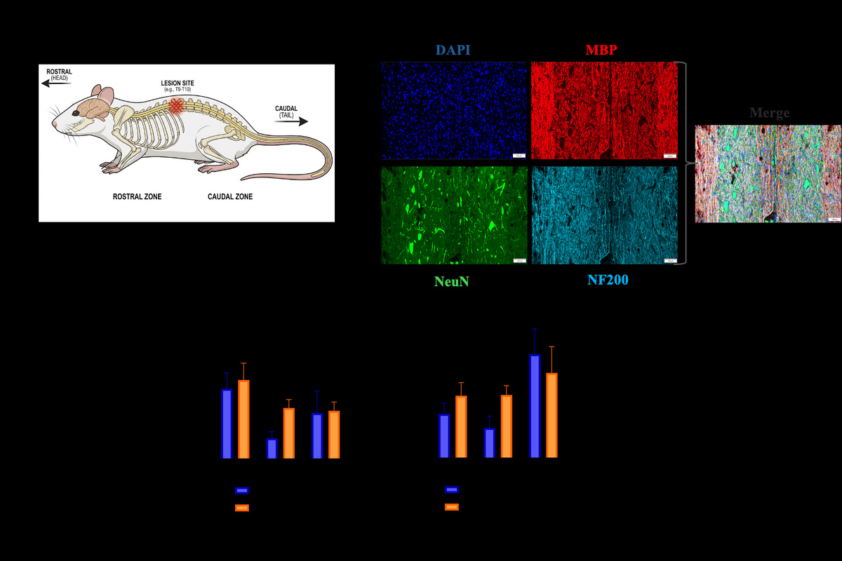
Two systems are commonly used to describe a traumatic spinal injury. AOSpine classifies the shape of the damage to the bones and ligaments (types A, B and C, with C the most unstable). The ASIA scale grades neurological function: how much movement and feeling remains. This study asked whether the first tells you anything extra about outcome.

The study

Researchers looked at 366 people with traumatic spinal injuries at Bouaké Teaching Hospital in Côte d'Ivoire, using both retrospective data (2017-2021) and prospective data (January 2025 to June 2026). Injuries were about 35% type A, 22% type B and 43% type C. Data are from a paper in the Open Journal of Modern Neurosurgery; we've read the abstract summary, not the full paper.

What they found

AOSpine type matched mechanical markers well, such as dislocation, ligament injury and cord compression. But it wasn't independently linked to death (about 18%) or neurological recovery (41-52%). In the adjusted analysis, only the ASIA grade stayed independently associated with outcomes.

What it means

AOSpine describes how unstable the injury is, which matters for surgery, but doesn't add prognostic value beyond ASIA grade. The authors say both should be recorded together.

Limits and Q&A

It's a single-centre study, partly retrospective, in a low-resource setting where transport delays and limited care may affect outcomes. Data from Africa are scarce, which is part of its value.

Does this mean the fracture type doesn't matter? No. It matters for surgical decisions. It just didn't add to prediction here.

Why does African data matter? Most SCI research comes from high-income countries, so patterns may differ elsewhere.

Read full article → (external link)
Research Neuroscience 28 SEPT 2026

How Spinal Stimulation Talks to the Breathing Circuits After Neck Injury

A rat study in Communications Biology examines what cervical epidural stimulation actually activates in the breathing system after injury, and finds that stimulation timed to the breath works best.

Read More

Deep Dive

Breathing problems are the leading cause of death after spinal cord injury. Existing breathing pacers largely override the brain's own rhythm. Researchers would like stimulation that works with it instead, and this study asks a basic question: when you stimulate the spinal cord in the neck, what are you actually switching on?
Why breathing is different from walking

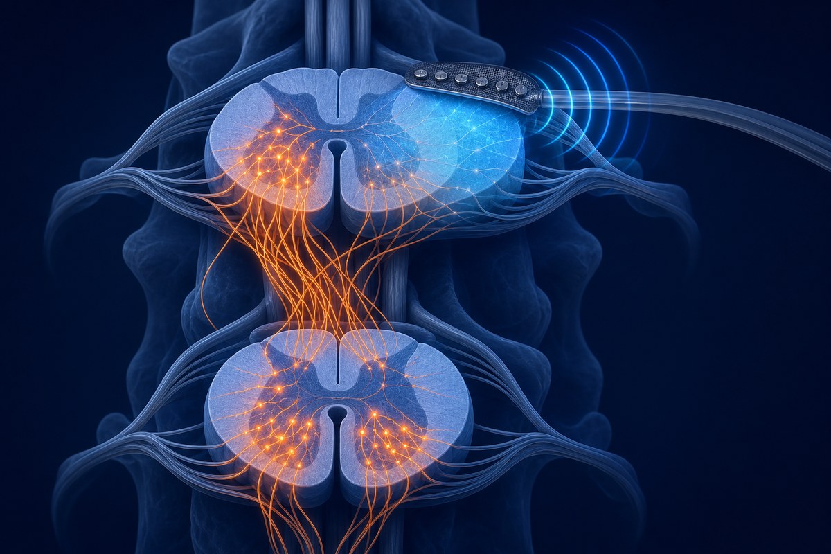
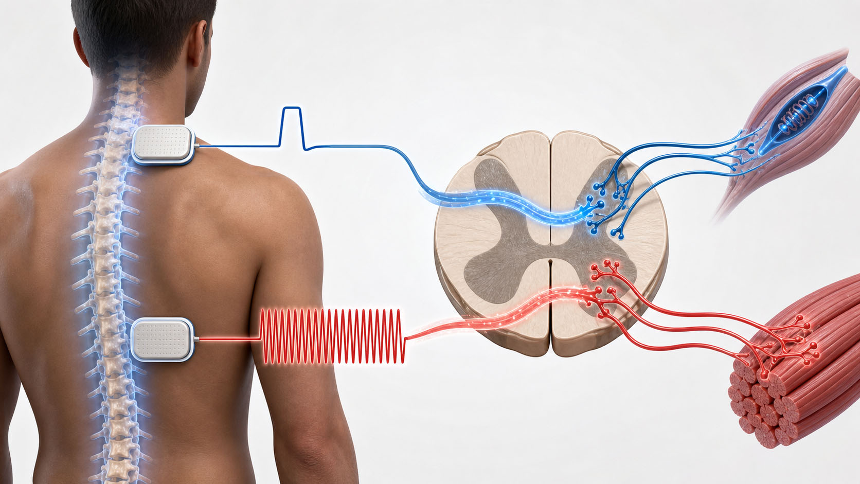
Epidural stimulation (electrodes placed on the outer surface of the spinal cord) has helped people move their legs, because the spinal cord holds its own movement rhythm generators, called central pattern generators. Breathing is different: its rhythm generator sits in the brainstem, not the spinal cord. So it wasn't clear what stimulating the cord in the neck was recruiting.

What the researchers did

In anaesthetised rats with a neck-level (cervical) spinal cord injury, the team compared three stimulation patterns: triggered on the breath in, triggered on the breath out, and constant (tonic) stimulation. They recorded output from the diaphragm, the main breathing muscle, and looked at markers of recently active cells (immediate early gene expression).

What they found

All three patterns made the diaphragm fire. But getting a properly shaped breathing pattern needed two things: signals descending from the brain and inhibition within the spinal cord. Fast responses and rising gene activity suggested stimulation was recruiting local spinal interneurons (relay cells within the cord), including inhibitory ones.

The authors conclude that stimulation patterned to the breath in is important.

Caveats

This is an animal study in anaesthetised rats, and it explains mechanism rather than testing a treatment. It doesn't show that any approach improves breathing in people.

Reader Q&A

Is this a breathing treatment I could get? No. It's early mechanistic research that could help design better stimulators later.

Why does timing to the breath matter? Stimulation that fits the body's own rhythm seems more likely to produce natural breathing than stimulation that overrides it.

What are interneurons? Cells inside the spinal cord that pass signals between other neurons. Here, some of them act as brakes that shape the output.

Read full article → (external link)
Research Brain-Computer Interface 28 SEPT 2026

BrainGate Study: Watching a Hand and Trying to Move Engage the Brain Differently

Brain recordings from two people with tetraplegia showed stronger responses to more human-like virtual hands during observation, but similar responses across shapes during attempted movement. No real robot was controlled.

Read More

Deep Dive

How much should a brain-controlled robotic gripper look like a human hand? A BrainGate study in PNAS explored one piece of that question by recording motor-cortex activity (the brain area that plans movement) from two people with tetraplegia (paralysis affecting all four limbs) while they watched virtual grasping movements. Brown University reported it on 23 September 2026.

Watching different shapes

Both participants have tiny electrode arrays implanted in the motor cortex as part of the BrainGate trial. They watched animations of grasping by a realistic human hand, a human-shaped robot hand, a three-pronged claw and a cube. Motor-cortex activity was strongest for the most human-like shapes and decreased as shapes became less human. This appeared both across the recorded population and in individual neurons.

In a separate set of animations, moving dots could be read as a hand. When one participant recognised that the dots represented a hand, activity rose. This suggests understanding matters, but it comes from a single participant.

Trying to move

When the participants attempted the grasp themselves while watching, responses were similar for every shape. The researchers suggest that the signal of trying to move can override differences caused by what is on screen. That raises a design question: a useful gripper might not need to look like a hand to work with a brain-computer interface. The study did not test that.

What about "mirror neurons"?

Mirror neurons are cells said to respond both when you act and when you watch an action. Lead author Jacob Gusman says the observation activity is not driven by specialised mirror-neuron cells but reflects wider network effects. Senior author Leigh Hochberg says the findings will help inform the next generation of implantable interfaces.

Limits

Two participants watching virtual animations cannot tell us which gripper gives better control, or help with daily tasks. Testing real devices is the next step.

Reader Q&A

Does this mean brain implants can control any robot hand? Not yet shown. This was recordings during observation, not device control.

Why do the different shapes matter? Designers need to know whether what a person sees interferes with the signals a decoder reads.

Is this a treatment? No. It's basic research to improve future interfaces.

Read full article → (external link)